🤖 Generated with AI assistance · Does not replace individualized medical advice.
DeepLNM: Multimodal AI Predicts Lymph Node Metastasis in Colorectal Cancer
AI Transparency Notice: This post was generated by an AI assistant to summarize and contextualize published scientific literature. It is intended for educational and informational purposes only and does not constitute medical advice, diagnosis, or treatment. Always consult a qualified healthcare provider for clinical decisions.
Lead
DeepLNM, a multimodal foundation model integrating histopathology, CT imaging, and clinical text, demonstrates robust performance in predicting lymph node metastasis (LNM) in colorectal cancer. With a prospective AUC of 0.845 PMID: 42873913, it offers a potential tool to reduce diagnostic errors and support staging accuracy.
Key Facts
| Metric | Value | Context |
|---|---|---|
| Internal AUC | 0.922 | Multi-center training cohort PMID: 42873913 |
| External AUC | 0.872 / 0.848 | Two independent external validation cohorts PMID: 42873913 |
| Prospective AUC | 0.845 | Registered clinical trial (NCT07239063) PMID: 42873913 |
| User Study Impact | 95.0%–97.3% | Diagnostic accuracy for junior pathologists PMID: 42873913 |
Why It Matters / Context
Accurate assessment of lymph node metastasis (LNM) is the cornerstone of prognostic stratification and treatment selection in colorectal cancer (CRC). Standard-of-care relies on surgical pathology, yet imaging and routine histological examination can yield false-negative or false-positive results, leading to inappropriate adjuvant therapy or unnecessary surgery.
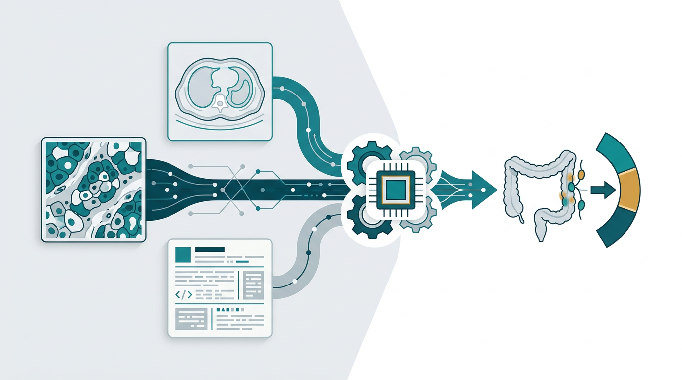
The development of DeepLNM addresses a critical gap in digital pathology: the integration of multimodal data. While previous AI models often focused solely on whole-slide images (WSI) or radiology, DeepLNM combines H&E-stained WSIs, CT scans, and structured clinical text. This approach mimics the holistic decision-making process of an experienced oncologist, who considers morphology, anatomical context, and patient history simultaneously.
The study’s significance lies not only in its high predictive accuracy but also in its rigorous validation strategy. By moving beyond retrospective analysis to include external cohorts and a prospective clinical trial, the authors provide stronger evidence for clinical utility. Furthermore, the inclusion of a user study highlights the model’s potential to augment human expertise, particularly for junior pathologists, thereby addressing workforce challenges in diagnostic pathology.
Details
The study evaluated DeepLNM across multiple cohorts to assess generalizability and clinical impact. The model was trained on a multimodal dataset from three hospitals, featuring matched H&E-stained whole-slide images, CT scans, and clinical text.
Performance Across Cohorts
| Cohort Type | AUC | Notes |
|---|---|---|
| Internal (Training) | 0.922 | Multi-center foundation model training set PMID: 42873913 |
| External 1 | 0.872 | Independent validation cohort PMID: 42873913 |
| External 2 | 0.848 | Independent validation cohort PMID: 42873913 |
| Prospective | 0.845 | Registered trial NCT07239063 PMID: 42873913 |
| Gastric Cancer | 0.793 | Cross-cancer generalizability test PMID: 42873913 |
User Study Outcomes
In a controlled user study, DeepLNM was used as a decision-support tool. The results indicated a significant improvement in diagnostic accuracy, particularly among junior pathologists, with accuracy rates ranging from 95.0% to 97.3% PMID: 42873913. Additionally, the model reduced diagnostic time and increased user confidence in their assessments.
Interpretability
To address the “black-box” concern inherent in deep learning, the authors utilized explainable AI techniques to identify specific histopathological features associated with LNM. This transparency is crucial for gaining pathologist trust and regulatory acceptance, as it allows clinicians to verify that the model is focusing on biologically relevant regions rather than artifacts.
Limitations
Critical Appraisal:
* Retrospective Bias in Training: While the prospective cohort is a strength, the initial training and internal validation were retrospective. This may introduce selection bias that does not fully reflect real-world clinical workflows.
* Cross-Cancer Performance Drop: The AUC dropped to 0.793 in the gastric cancer cohort PMID: 42873913, suggesting that while the model has some generalizability, it is not yet a universal tool for all gastrointestinal cancers without retraining or fine-tuning.
* User Study Scope: The user study focused on diagnostic accuracy and time but did not report on long-term clinical outcomes (e.g., survival, recurrence) directly attributable to the AI-assisted diagnosis.
* Data Heterogeneity: The study relies on data from three hospitals. Variability in staining protocols, CT scanner models, and data annotation standards across these sites could impact the model’s performance in other healthcare systems.
Clinical Takeaway
Clinicians should view DeepLNM as a promising decision-support tool that may enhance staging accuracy and reduce diagnostic variability, particularly for less experienced pathologists, but it should not replace expert pathological review without further prospective, outcome-based validation.
Full Reference
Zhang Z, Lu C, Wang Z, Cen Z, Zhou Y, Li D. DeepLNM: A Multimodal Model for Predicting Lymph Node Metastasis in Colorectal Cancer. Advanced Science (Weinheim, Baden-Wurttemberg, Germany). 2026. PMID: 42873913 [DOI: 10.1002/advs.77860]