🤖 Generated with AI assistance · Does not replace individualized medical advice.
AI Act Transparency Notice
This content was generated with the assistance of artificial intelligence. It is intended for informational and educational purposes only and does not constitute medical advice, diagnosis, or treatment. Always seek the advice of a qualified health provider with any questions regarding a medical condition.
CIPHER: A Deep Learning Foundation Model for Predicting ICI-Induced Pneumonitis in NSCLC
Key Facts
| Metric | Value | Context |
|---|---|---|
| External Validation AUC | 0.83 | Superior to radiomics (DeLong p=0.0318) PMID: 42759982 |
| Pretraining Dataset | 590,284 CT slices | From 2,500 NSCLC patients PMID: 42759982 |
| External Cohort Size | 116 patients | 20 ICI-P cases; 96 controls PMID: 42759982 |
| Balanced Accuracy | 81.7% | External validation performance PMID: 42759982 |
Why It Matters / Context
Immune checkpoint inhibitors (ICIs) have transformed the treatment landscape for non-small cell lung cancer (NSCLC), yet their efficacy is often tempered by immune-related adverse events (irAEs). Among these, ICI-induced pneumonitis (ICI-P) is particularly concerning due to its potential severity and the difficulty in distinguishing it from tumor progression or infectious etiologies. Current risk stratification relies heavily on clinical factors (e.g., smoking history, autoimmune disease) and radiomics features, which often lack the sensitivity and specificity required for precise patient selection.
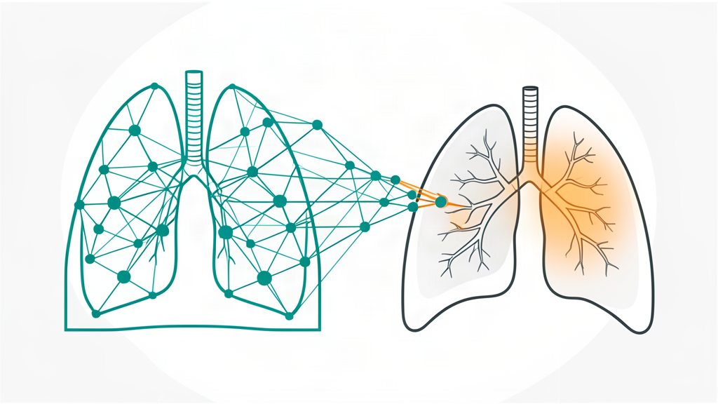
The development of CIPHER (Checkpoint-Inhibitor Pneumonitis Hazard EstimatoR) represents a shift toward “foundation models” in oncology imaging. By leveraging self-supervised learning on a massive dataset of 590,284 CT slices from 2,500 patients, the model learns robust representations of heterogeneous lung parenchyma without requiring extensive labeled data for pretraining PMID: 42759982. This approach addresses a critical gap: the ability to identify high-risk patients before ICI initiation, enabling closer monitoring and timely intervention. The study demonstrates that deep learning can extract subtle, sub-visual patterns from baseline CT scans that are predictive of future immune toxicity, outperforming traditional hand-crafted radiomics features.
Details
The study employed a rigorous multi-stage validation process, moving from internal development to external independent validation. The model was fine-tuned on a subset of the MD Anderson Cancer Center cohort and evaluated against clinical, radiomics, and ensemble comparator models.
Performance Comparison: Internal vs. External Validation
| Model Type | Internal AUC Range | External AUC | External Sensitivity | External Specificity | Notes |
|---|---|---|---|---|---|
| CIPHER (Deep Learning) | 0.77 – 0.85 | 0.83 | 80.0% (16/20) | 83.3% (80/96) | Superior specificity; robust classification PMID: 42759982 |
| Radiomics Model | N/A | N/A | 85.0% | 45.8% | High sensitivity but low specificity PMID: 42759982 |
| Clinical/Ensemble | N/A | N/A | N/A | N/A | Outperformed by CIPHER in head-to-head PMID: 42759982 |
Note: External validation cohort comprised 116 patients (20 ICI-P, 96 non-ICI-P). CIPHER correctly identified 16 of 20 ICI-P cases and 80 of 96 non-ICI-P cases PMID: 42759982.
The statistical superiority of CIPHER over the radiomics model in the external cohort was confirmed with a DeLong p-value of 0.0318 PMID: 42759982. While the radiomics model achieved a slightly higher sensitivity (85.0%), its specificity was markedly lower (45.8%), leading to a higher rate of false positives. In clinical practice, high specificity is crucial for risk stratification tools to avoid unnecessary intensive monitoring or treatment delays in low-risk patients. CIPHER’s balanced accuracy of 81.7% in the external cohort suggests a more reliable overall classification performance PMID: 42759982.
Limitations
Critical Limitations for Clinical Translation
1. Small Event Rate in Validation: The external validation cohort contained only 20 ICI-P cases out of 116 patients. This small number of positive events limits the statistical power to precisely estimate sensitivity and specificity, and confidence intervals for these metrics are likely wide.
2. Retrospective Design: The study is retrospective. While external validation was performed, the lack of prospective data collection means that real-world workflow integration, image quality variability, and clinician adherence to the risk score remain untested.
3. Single-Modality Input: CIPHER relies solely on baseline CT scans. It does not incorporate genomic data (e.g., PD-L1 status, TMB) or detailed clinical history, which may interact with imaging features to influence pneumonitis risk.
4. Generalizability Concerns: The model was pretrained on NSCLC patients. Its applicability to other cancer types treated with ICIs (e.g., melanoma, renal cell carcinoma) is unknown and requires further investigation.
5. Lack of Prospective Validation: The authors explicitly state that CIPHER “warrants prospective validation before clinical translation” PMID: 42759982.
Clinical Takeaway
Clinicians should view CIPHER as a promising, non-invasive adjunct for pre-treatment risk stratification in NSCLC, but it should not yet replace standard clinical judgment or prospective monitoring protocols until validated in larger, multi-institutional prospective trials.
Full Reference
Muneer A, Showkatian E, Kitsel Y, Saad MB, Sujit SJ, Soto F. CT-based deep foundation model for predicting immune checkpoint inhibitor-induced pneumonitis risk in lung cancer. Journal for Immunotherapy of Cancer. 2026. PMID: 42759982 [DOI: 10.1136/jitc-2026-015014]